
ContentOptions To A Ct Scan.Radiologists Sharing Ct Scans, X.What Happens Throughout My Exam.What Ar
Se næste indlæg >
what are the differences between medical scan types
remingtonrrze
![]()
Content
- Imaging Plays A Vital Role In Modern-day Medication Modern-day Imaging Methods.
- When A Ct Scan Is Utilized.
- Specific Indicators For Cranial Ultrasound:.
- What Happens During My Evaluation.
Commonly, the cartilage in among the joint compartments is most drastically influenced. The standing X-rays may show narrowing of the involved joint room of the knee. Modified placement of the knee joint is really common as either the cause or as a result of osteoarthritis.
You need to discuss this with your referring physician and tell us at the time of scheduling if you assume it will certainly be of advantage. Throughout a cephalogram procedure, you will certainly be asked to stand or being in front of the x-ray maker as well as little secures will certainly be positioned in your ears to help align your head. Prior to the x-ray you will be asked to eliminate any kind of jewelry or eyeglasses. You will after that be required to an x-ray space, where a professional will certainly offer you directions and also perform the treatment. Our payment mirrors the expense of delivering exclusive radiology and also industry-leading person care.
Imaging Plays A Crucial Duty In Modern Medication Modern-day Imaging Techniques.
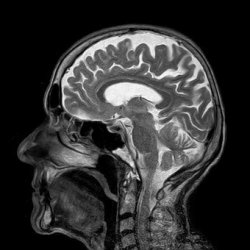
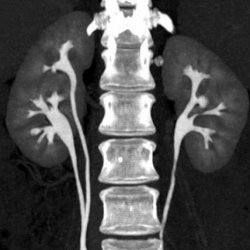
Clinical imaging such as X-rays, ultrasounds as well as CT scans can help with diagnosis, therapy, and monitoring of several medical conditions, from fundamental bone fractures to bust cancer. But both main points you ought to consider prior to taking on these procedures are the dangers included from the direct exposure to radiation, and also what it's going to cost you. A Computed Tomography scan usages numerous x-rays to generate cross-sectional layers of the body. This provides a much more 3 dimensional sight of the client's body compared to a straightforward x-ray. Although a CT scan is quick as well as provides much more detail than an x-ray, it likewise provides an enhanced quantity of exposure to radiation. CT scans are frequently used to identify embolism, cancer cells, interior bleeding and also more intricate photos of damaged bones. Magnetic Resonance Imaging uses a magnetic field as well as radio waves to show in-depth images of body organs, bones, ligaments and also soft cells.
Utilized for medical diagnosis to show information of components inside your body, such as the lungs, brain, stomach organs, bones and also capillary. Nuclear medication and PET DOG scans utilize a percentage of contaminated product. This is either infused right into you, or you breathe it in or swallow it. A special cam is after that made use of which finds the energy from the radioactive product in your body. Some imaging procedures utilize X-rays which are a special kind of radiation called 'ionising radiation'.
When A Ct Scan Is Made Use Of.
GP-referred imaging is largely paid for by Medicare, which has an arranged charge for imaging services as well as supplies a repayment each. Clients may additionally require to add directly when there is a gap between the Medicare discount and the quantity charged by the supplier, or in uncommon instances, where no Medicare rebate exists. If a supplier is prepared to accept the discount as complete settlement, they can bulk costs Medicare, cutting the individual out of the x ray clinic near me payment procedure as well as reducing both management and accounting prices.
Within this website, the relative radiation level of each imaging examination is presented as below. The LNLT design indicates that no dosage of IR, nevertheless little, is entirely without threat. This design estimates the ordinary life time risk of induction of a deadly cancer cells from exposure to 5 milliSieverts to be approximately 1 in 4000 and that to 20 mSv to be 1 in 1000. The threat is substantially higher than standard in children and young people and lessens with age over the age of 40 years.
Details Indicators For Cranial Ultrasound:.
how the report is communicated from the practice or medical facility to your doctor (i.e. phone, email, fax or mail). Radiographers are educated to utilize the tiniest possible amount of X-rays needed to generate an adequate image. The whole process is uncomplicated, and you will certainly not feel anything weird or feel any kind of various throughout the exam. It is important that you stay entirely still when the radiographer instructs you to, as any type of movement might produce a blurred image.
- CT scanning tools includes a huge gantry with a circular opening.
- Simple radiography/X-rays are executed in the analysis imaging department of the majority of healthcare facilities (although this depends on the size of healthcare facility, as some little health centers do not have X-ray facilities).
- X-rays are made use of to take pictures of bones as well as some components of within your body, including the lungs.
- Inside the gantry is a turning ring that lugs the x-ray resource and also digital x-ray detectors.
- Radiographers and radiologists work carefully with each other in private clinical centres, significant public and exclusive hospitals and specialist centers, such as cancer facilities.


Ingen kommentarer endnu