
ContentWhat Takes Place Throughout My Exam.Ct Scan.Which Is A Lot More Efficient: Mri Or Ct?Ct.Guide
Se næste indlæg >
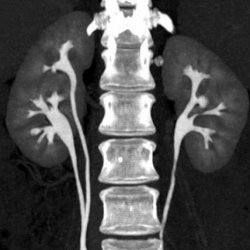
ct scan
remingtonrrze
Content
- So, Which Is Much More Efficient?
- Analyzing The Danger.
- Locate A Doctor.
- Typical Effective Doses Of Imaging Investigations.
- ' A Put In The Face': Facebook Ban On Australian Information Can Have Major Results In The Pacific.
Have I taken a history, carried out a physical exam as well as pertain to a provisional clinical medical diagnosis? The significance of the outcome of a test can not be accurately assessed without a pre-test chance of the disease being tested for. In Spain, Dr Edgar Lorente shared his cases to offer others an instance of how the condition can search in much less modern photos. Radiopaedia functions like Wikipedia, crowdsourcing information on illness and amassing a substantial archive of photos which can be accessed by anyone.
This is specifically helpful when taking a look at soft tissue for irregularities, or detecting problems in these intricate areas. When booking for a CT evaluation, please suggest the assistant if you have had a previous allergic reaction to x-ray contrast. Relying on the medical examination, the CT scan may last anywhere from several minutes to over half a hr. The examination table slides Brighton Radiology Melbourne into a round opening in the machine while numerous pictures are caught. is a great advent in the medical diagnosis of cardiac conditions, as its high resolution as well as high speed allows for clear coronary artery pictures.
So, Which Is A Lot More Reliable?
Almost every client that is referred to an Orthopaedic surgeon for a hip or knee issue ought to have an X-Ray. This is NOT as useful as seeing the real photos themselves due to the fact that Orthopaedic specialists are searching for certain things that radiologists might not understand. In recap, if the prospective benefit of the scan outweighs the risk, after that the scan is justified. If the patient requires a scan for therapy or administration then they must not be postponed having one. The danger of cancer cells induction by IR is a deferred danger that might happen from 5 to 15 years after direct exposure.
![]()
It is not required for arthritis or any soft cells problems such as meniscal tears or ligament tears. A CT Scanogram is of some minimal benefit however a scientific evaluation is often more useful. CT scans are also helpful considering patellar tracking, in which situation an unique request requires to be gotten looking at different degrees of flexion, normally from 0-45 levels. Normal comply with up X-Rays are essential once a client has had a joint replacement. The specialist looks at alignment, the sort of joint, wear, the knee, loosening of the prosthesis or any loose bodies. Generally, people should have check X-Rays each or two years depending on the type of joint and the age of the person. Both 3D and 4D ultrasounds give three-dimensional images of the fetus, revealing more structural as well as inner details from various angles.
Assessing The Risk.
The X-ray resource and also a detector after that rotate around the patient creating a slim 'fan-shaped' beam of light of X-rays that goes through a section of the client's body to create a snapshot. In a suitable world, we 'd have the ability to identify as well as treat people with no damaging negative effects. Clinical imaging remains one of the very best means to achieve that goal, being able to see what's taking place inside the body without the need for surgery or other invasive procedures.
The standard x-ray uses ionising radiation to produce images of the body's inside. Going an action additionally, the CT scan is an x-ray maker that turns, creating cross-section scans of the body. When these scans are combined with each other, a 3D image is established that can offer useful understanding as well as info to medical professionals and radiologists.
Find A Doctor.
Magnetic resonance imaging includes a big apparatus which contains a high-strength magnet. This magnet surrounds a system on which a person lies throughout the examination. MRI creates cross-sectional, multiplanar photos that allow the orthopedist to assess anatomic frameworks– including different kinds of cartilage, tendons, tendons, and bones. Imaging modern technologies made use of at HSS to identify hip discomfort consist of X-ray, calculated tomography, magnetic resonance imaging, andultrasound. The orthopedist determines which technique to use, depending upon the believed disease or injury, the details type of info required as well as, in many cases, the age or basic problem of the individual.


Ingen kommentarer endnu